Full Mouth and Intraoral X-ray Examination
In a typical routine dental examination, only the surface of the teeth or shallow gum health can be observed. In order to detect conditions such as root abnormalities, inflammation of the dental pulp, or internal tooth decay (cavities) within the teeth and jawbone, X-ray imaging is necessary.
X-ray imaging assists dentists in diagnosing whether patients have cavities, following up on root canal treatments, examining periodontal disease for gum and alveolar bone damage, and discovering hidden supernumerary teeth, skeletal diseases, etc., enabling patients to be diagnosed early and receive treatment.
In addition to helping dentists provide timely diagnosis and treatment for patients, X-ray imaging can also serve as a basis for evaluating future changes in teeth. X-ray diagnosis is part of a comprehensive oral examination, and depending on the patient’s oral condition, dentists will use different X-ray imaging technologies.
Panoramic Radiograph (OPG)
Panoramic Radiograph, abbreviated as OPG, is also known as a full-mouth dental X-ray or a full-mouth dental radiograph. It is a method of taking X-ray images of the entire oral cavity. A professional camera is used for taking full-mouth X-rays, providing a wider field of view on a single film, allowing multiple areas and structures within the oral cavity to be examined at once.
Compared to traditional small intraoral X-rays, panoramic radiographs can display oral structures more comprehensively, including the jawbone, temporomandibular joints, teeth, tooth roots, occlusal relationships, jawbone tumors, and other information. Therefore, panoramic radiographs can provide more comprehensive and detailed images of oral structures, helping dentists make more accurate diagnoses and treatment plans for patients' oral issues.
Panoramic radiographs are commonly used for diagnosing conditions before tooth extraction, prior to installing dental crowns, before oral surgery, and for oral cancer screening. The information about jawbone structure and temporomandibular joint conditions in panoramic radiographs is crucial for procedures such as tooth extraction, dental implants, orthodontic treatments, and other oral therapies.
Periapical X-rays and Bite-wing X-rays
Periapical X-rays and Bite-wing X-rays are two common types of dental X-ray images used to examine the condition of teeth and surrounding tissues.
1. Periapical X-rays:These X-rays are used to examine the entire tooth, including the root and surrounding tissues. They show the apex (tip) of the tooth and help detect infections around the root apex, damage to the tooth root, or other issues.
2. Bite-Wing X-rays:These X-rays are used to examine the biting relationship between teeth, primarily showing the occlusal surfaces of the teeth and gum condition. They are commonly used to detect cavities between teeth, dental plaque around teeth, and dental calculus.
In summary, Periapical and Bite-wing X-rays are primarily used for localized examination of individual teeth or specific areas, while panoramic radiographs provide a more comprehensive oral examination. Depending on the condition and the dentist's recommendations, different types of dental X-ray images can be used to aid in the diagnosis and treatment of oral issues.
Cone Beam Computed Tomography (CBCT)
Cone Beam Computed Tomography (CBCT) is a specialized type of X-ray equipment that allows for clear examination of the internal structures of bones and teeth. Through 3D scanning, CBCT can assess the morphology of teeth, soft tissues, nerve pathways, and bones. Compared to traditional computed tomography scans, CBCT generates less radiation and provides higher accuracy.
When undergoing a CBCT scan, there is no special preparation required. However, patients should inform the dentist early if they are pregnant or have any allergies. During the CBCT scan, patients should wear loose, comfortable clothing and remove any jewelry.
CBCT scans offer many advantages. The focused X-ray beam reduces scatter radiation during the scanning process, thereby improving image quality. A single scan can produce multiple views and angles for a more comprehensive assessment. Cone beam CT scans provide more information than traditional dental X-rays, aiding in the development of more precise treatment plans. The CT scanning process is painless, non-invasive, and accurate, capable of displaying images of both bones and soft tissues. After a CT examination, there is no residual radiation left in the patient's body.
Common Questions about CBCT (Cone Beam Computed Tomography):
What Are The Benefits Of CBCT Scanning?
- The focused X-ray beam reduces scatter radiation, thereby enhancing image quality.
- A single scan generates multiple views and angles for a more comprehensive evaluation.
- CBCT provides richer information than traditional dental X-rays, aiding in the development of more accurate treatment plans.
- The scan is painless, non-invasive, and accurate.
- It can display images of both bones and soft tissues simultaneously.
- After a CBCT examination, there is no residual radiation left in the patient's body.
Why Is A CBCT Scan Necessary?
A CBCT scan is necessary because it can display detailed images of bones, nerves, and soft tissues, allowing dentists to accurately diagnose a patient's oral health condition before surgery and develop more precise treatment plans. It is important to note that CBCT scanning is crucial for planning and successfully executing dental implant surgery.
Can CBCT Scans Detect Tooth Infections?
CBCT can help identify abscesses in painful teeth. Dentists can also use this technology to determine if an infection has spread, leading to abscesses in other areas. If the infection has spread to other areas in the neck, CBCT scans can be used to assess the extent of the infection.
What Is The Difference Between Ct And CBCT?
CBCT scanners use cone-shaped beam radiation from an X-ray source that covers a larger volume and rotates around the patient's head. In contrast, traditional CT scanners use a rotatable high-output anode X-ray tube, while CBCT scanners use low-power medical fluoroscopy tubes.
What Preparations Are Needed Before And After A CBCT Examination?
The preparation for a CBCT examination is simple. There is no special preparation required. Before the examination, you may need to remove items that could affect the images, such as jewelry, glasses, hairpins, and hearing aids. The CBCT examination is painless, and after completion, you can resume normal activities.
Digital 3D Tooth Impressions - Intraoral Scanner (iTero Intraoral Scanner)
The 3D intraoral scanner can scan a patient's mouth in real-time, generate 3D images, and has a close-range infrared function that can be used to detect cavities. This helps dentists explain dental issues to patients more easily and improves diagnostic accuracy.
In addition to being a dental examination tool, this advanced 3D scanner can replace traditional plaster impression techniques. The entire impression process only takes 5 minutes, which is 50% faster than traditional techniques, reducing the need for reprinting due to errors, and patients can also reduce discomfort caused by impressions.
Features Of Digital Dental Scanning:
- Real-time simulation of orthodontic results.
- Preview of simulated orthodontic treatment progress and the difficulty of correcting teeth.
- Impression process only takes 5 minutes.
- Fast and comfortable process.
- No radiation involved.
- Does not cause throat sensitivity or reflex vomiting as traditional impressions do.
- Suitable for various dental services including teeth straightening, implants, and braces.
To ensure oral health, regular exams are essential. Common dental problems including cavities, gum disease, poor breath, and uneven tooth colour have a big influence on appearance, self-esteem, and interpersonal interactions in addition to their negative effects on oral health.
A yearly examination may help maintain your teeth healthier, stronger, and free of discomfort in addition to preventing dental disorders including cavities and periodontal disease. You may save future dental care costs and time commitments by detecting problems early and getting treatment quickly.
Preventing oral diseases requires knowledge of proper oral hygiene steps and strategies. Knowledge of personal care is highly valued by our dental team. In order to help you develop better oral hygiene habits and advance oral health, our dentists and healthcare professionals answer your oral health-related concerns, offer advice on how to clean your teeth, and show appropriate brushing methods at each routine check-up.
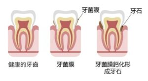
Gingivitis, periodontal disease, and bad breath could all be avoided by getting your teeth cleaned regularly. The goal of teeth cleaning is to return the teeth to their original clean and smooth condition by removing plaque, tartar, and stains that build up on the tooth's surface and in between the teeth.
Following a meal, food particles may get stuck to our teeth's surface or in between them. Even a brush won't get rid of this plaque easily. Bacterial activities can cause plaque to develop over time and possibly calcify into tartar. Bad breath and periodontal disease are among the consequences that can arise from tartar's uneven surface, which facilitates bacterial attachment to teeth. Gingivitis and periodontal disease may be avoided by cleaning your teeth using ultrasonic technology, which removes plaque and tartar.
Steps for Scaling and Polishing
1. The dentist will completely remove plaque and tartar from your teeth using an ultrasonic cleaning tool.
2. The dentist will next polish and smooth the tooth surfaces, maintaining the smoothness of the tooth surface and strengthening the outer layer of the teeth. This lessens the chance of developing periodontal disease and helps stop debris from building up on uneven surfaces.
Tips for Scaling and Polishing Aftercare
- In the days after having your teeth cleaned, you can feel a little uncomfortable and have gum bleeding in your mouth. Patients who have periodontal disease are more likely to have swelling and bleeding. Gum bleeding and soreness will eventually stop and the gums will recover back to normal with proper dental hygiene.
- Inflamed and swollen gums following tooth cleaning may diminish when the irritation goes down. Gum recession can lead to tooth sensitivity, causing discomfort when eating cold, hot, sweet, or sour foods. Desensitising toothpaste can be used as needed to ease the discomfort. It is recommended that you get in touch with our healthcare professionals immediately if the sensitivity continues.
FAQ
Why Is Regular Dental Cleaning Essential?
The interdental space is the small area that exists between our teeth and gums when there isn't a complete seal. Food particles may get stuck in this area after eating, making it challenging to clean even with frequent brushing. This can result in the development of plaque, which sticks to the gums and tooth surfaces. Plaque is progressively hardened into tartar by the metabolic processes of bacteria.
Tartar begins as a thin, white deposit but hardens soon to develop a dark green or brown colour. The formation of tartar makes it more difficult to get rid of the bacteria and plaque that irritates the gums and causes periodontal disease.
Gum recession, bone loss surrounding teeth, tooth loosening, and eventual tooth loss can all be symptoms of severe periodontal disease.
Using dental equipment to remove plaque and tartar from the surfaces of the teeth and the spaces between them, teeth cleaning aims to stop the growth of bacteria and prevent consequences including gum disease, periodontal disease, and bad breath. Along with checking for any more potential dental health problems, the dentist can address them appropriately.
Is Dental Cleaning Necessary?
For the maintenance of oral health, dentists advise routine dental cleanings. Although people should generally get their teeth cleaned every six to nine months, the following people are more vulnerable to periodontal disease and should get their teeth cleaned every three to six months:
- Smokers.
- Denture wearers.
- Those who have undergone or are undergoing orthodontic (braces) treatment.
For patients with periodontal disease, dentists will recommend an increased frequency of dental cleanings based on the severity of the disease.
What Are The Benefits Of Dental Cleaning?
- Removing plaque and tartar adhered to the tooth surface and in between teeth.
- Allowing dentists to diagnose and treat cavities or other dental diseases early.
Does Dental Cleaning Damage Tooth Enamel?
In general, dental cleaning does not harm the enamel of the teeth
In order to efficiently remove both tartar and plaque during a dental cleaning, dentists use ultrasonic vibrating devices that cause water molecules to vibrate and contact the tartar. This allows the tartar to come off.
During A Dental Cleaning, Why Does The Dentist Use Tools To Scrape My Teeth?
In most cases, dentists do not need to use dental instruments to directly scrape off tartar during a routine dental cleaning. Only in severe cases of periodontal disease where tartar has built up deep below the gum line, and the patient requires periodontal treatment, dentists may use a periodontal scaler to clean the tooth root surfaces and remove tartar under the gums to eliminate periodontal pockets.
Regular dental cleanings are advised to maintain the cleanliness of teeth and gums as the best way to protect your teeth.
Can Dental Cleaning Cause Gaps Between Teeth To Widen Or Loosen Teeth?
Undergoing a dental cleaning treatment does not cause gaps between teeth to widen or teeth to loosen.
Generally, the widening of gaps between teeth and loosening of teeth primarily occur in patients with severe periodontal disease. When a patient has severe periodontal disease, tartar buildup can lead to bacterial infection, causing gum inflammation and gradual recession, affecting the surrounding alveolar bone, eventually leading to loosening of teeth. However, during a dental cleaning treatment, the dentist removes the tartar that fills the gaps between teeth, helping to alleviate gum inflammation symptoms, rather than causing gaps to widen or teeth to loosen.
Additionally, improper use of toothpicks for cleaning or age-related gum and alveolar bone degradation can also contribute to issues with gaps between teeth. It is recommended to use interdental brushes to thoroughly clean the gaps between teeth, prevent tartar buildup, and help maintain oral health.
In conclusion, dental cleaning treatment itself does not cause the widening of gaps between teeth or tooth mobility. Instead, it helps alleviate symptoms of periodontal disease and improve overall oral health.
Is It Because The Dentist Is Too Forceful When Cleaning My Teeth If There Is Bleeding During Dental Cleaning?
Gum bleeding should not typically occur from using adequate dental cleaning techniques. Bleeding during dental cleaning is usually a sign of periodontal disease, which causes swelling, inflammation, and irritation in addition to making the gums prone to bleeding.
The primary goal of dental cleaning is to minimise inflammation in the gums by clearing tartar from the spaces between teeth.
Can Dental Cleaning Whiten Teeth?
Generally speaking, undergoing a dental cleaning does not guarantee teeth whitening results.
Healthy teeth are often slightly yellow in colour, and plaque buildup on the surface of the teeth can make them look even more yellow. In addition, smoking, drinking coffee or tea for an extended period of time, and other habits can cause pigment deposits on the surface of the teeth, which darken the appearance of teeth.
The main purpose of dental cleaning is to thoroughly remove yellowish plaque on the tooth surface and tartar formed. As a result, it's common for many people to notice that their teeth look somewhat brighter following dental cleaning.
What Should Be Paid Attention To After Dental Cleaning?
- After dental cleaning, you may experience tooth sensitivity, which typically subsides gradually within two to three days.
- For patients with periodontal disease, gum swelling may gradually decrease after dental cleaning, leading to increased tooth sensitivity. Patients may experience discomfort when eating cold, hot, sweet, or acidic foods. In such cases, patients can consider using desensitizing toothpaste to alleviate discomfort.
- Patients with periodontal disease may experience mild gum bleeding after dental cleaning. Continuing to maintain oral hygiene will gradually improve oral discomfort and gum bleeding, leading to the restoration of gum health.
What Is A Dental Hygienist? What Is The Difference Between Them And A Dentist?
A dentist, on the other hand, is a healthcare professional trained in dental medicine with the ability to diagnose oral diseases and provide oral treatments. Dentists can perform more complex oral surgeries, such as implant treatments, and other treatments, such as root canal treatments, including teeth cleaning treatment.
A dental hygienist is a licensed professional dental care provider recognized internationally. Under the guidance of a dentist, dental hygienists perform tasks such as teeth cleaning, removal of tartar, tooth polishing, taking X-rays, applying fluoride for cavity prevention, and providing oral health education tailored to individual patients to achieve the long-term goal of prevention over treatment.
In summary, dental hygienists focus on teeth cleaning, educating patients on oral hygiene and preventive care, while dental cleaning by a dentist refers to the teeth cleaning procedure performed by a dentist, who can also provide more complex oral treatments when needed.
The purpose of dental filling is to restore the appearance and function of a tooth, prevent further damage to the tooth, and alleviate pain. Professional tooth fillings are typically used to treat tooth damage caused by cavities or trauma. When the surface of a tooth is damaged, a dental filling procedure may be recommended by a dentist.
The Dental Filling Procedure Typically Involves The Following Steps:
- The dentist will examine the condition of all the patient's teeth to assess whether dental filling or other treatments are necessary.
- Depending on the individual patient's situation, the dentist may use local anesthesia to reduce pain.
- Subsequently, the dentist will use dental instruments to remove the damaged part of the tooth, ensuring cleanliness and sterility, and confirming the size and shape of the cavity.
- The cavity is filled with amalgam or composite material along with an adhesive.
- Finally, the dentist will polish the filling material to restore the appearance and occlusion of the tooth.
FAQ
What Are The Common Materials Used For Dental Fillings?
The common materials used for dental fillings include silver amalgam and composite resin.
Amalgam
Amalgam is an alloy made up of silver, tin, copper, and mercury. It is known for its durability, ease of use, and safety. Amalgam fillings are typically used for filling smaller cavities. However, due to their silver-gray color, they are less aesthetically pleasing and are primarily used in the back teeth to withstand greater biting forces.
Composite Resin
Composite resin has a color that closely matches the natural color of teeth and is less prone to plaque buildup. It is commonly used for cosmetic purposes, such as improving the shape, size, and color of teeth, or for filling cavities in visible areas like the front teeth where aesthetics are important.
How Long Does A Dental Filling Procedure Take?
The duration of a dental filling procedure can vary depending on factors such as the materials used and the depth of the cavity, but typically the entire filling process can be completed within 20 to 60 minutes. Some more complex cases may require multiple treatments.
Does Getting A Dental Filling Hurt?
During a dental filling procedure, the dentist will use local anesthesia to patients who need it, so you should not feel pain throughout the process.
Sometimes, the filling process may make the tooth more sensitive to stimuli, leading to discomfort.
After getting a dental filling, it may take a few days to a week for your teeth to adjust to the new filling material. This sensation is typically temporary and should gradually diminish over time. If any discomfort persists or worsens, it is advisable to consult your dentist.
What Is The Difference Between A Dental Filling And A Root Canal?
A dental filling involves repairing the surface of a tooth and filling a cavity to prevent further damage and restore the appearance of the tooth.
On the other hand, root canal treatment is an effective method for treating pulp and periapical diseases, aiming to save the affected tooth or root. Root canal treatment involves removing the pulp and necrotic tissue from the root canal, proper disinfection, filling the root canal to eliminate adverse stimuli to periapical tissues, prevent or promote the healing of periapical diseases, and thus preserve the affected tooth or root.
What Should Be Noted After Getting A Dental Filling?
After receiving a dental filling, your dentist will provide you with recommendations, such as the timing of eating and drinking, temporary avoidance of certain foods, and a prescription for pain relief medication. Typically, within the first three days after getting a filling, it is advised to avoid chewing on hard objects to prevent deformation or damage to the filling material.
After getting a dental filling, it is important to pay special attention to oral hygiene to prevent tooth decay. This includes brushing with fluoride toothpaste twice a day and using dental floss to clean between teeth. It is advisable to minimize consumption of sugary foods and to schedule regular dental check-ups and cleanings.
Tooth extraction is the process of removing a tooth that has severe issues or is not suitable for preservation, such as severely decayed or fractured teeth. Typically, the procedure does not require general anesthesia or surgery. However, if a tooth is impacted or located very close to a nerve, surgical extraction may be necessary.
Sometimes, during other dental treatments such as orthodontic procedures, a dentist may recommend tooth extraction. Tooth extraction can be categorized into non-surgical and surgical methods, each with its own applications and treatment procedures.
Procedure for Non-Surgical Tooth Extraction:
- Oral Examination: The dentist will conduct a detailed examination of your mouth and teeth and may arrange for dental X-rays to assess the tooth's position, length, and the condition of the surrounding alveolar bone to determine the most suitable extraction method.
- Local Anesthesia: The dentist may use local anesthesia around the tooth area to ensure you do not feel discomfort or pain during the extraction.
- Extraction: The dentist will use a dental elevator to loosen the tooth and then extract it using forceps.
- Post-Extraction Care: After the tooth is removed, you may be instructed to bite down on a piece of gauze for 30 minutes to an hour to control bleeding.
It is essential to follow your dentist's post-extraction care instructions to promote proper healing and prevent complications.
FAQ
When Is Tooth Extraction Necessary?
Cases where tooth extraction may be necessary include:
- Severe periodontal disease leading to bone loss and tooth mobility
- Improper tooth positioning causing damage to surrounding tissues and infection
- Teeth that are too damaged by severe decay or fracture to be repaired
- Abnormal tooth appearance or structure
- Supernumerary teeth (extra teeth)
- Necessary for orthodontic treatment or other dental procedures
- Pathological reasons such as teeth associated with tumors
What Are The Differences Between Surgical And Non-Surgical Tooth Extraction?
Non-surgical tooth extraction
Non-surgical tooth extraction is typically used for simpler cases where the dentist believes the tooth can be removed without surgery.
The dentist will administer local anesthesia at the extraction site to ensure painlessness and then use dental instruments to remove the tooth. The process does not involve surgery or sutures.
Surgical Tooth Extraction
Surgical tooth extraction is typically performed in more complex cases, such as:
- The tooth is severely decayed and cannot be removed through a simple (non-surgical) extraction.
– Impacted wisdom teeth.
– The tooth is located close to a nerve, posing a risk of nerve injury.
– The tooth is deeply embedded in the jawbone, or the jawbone is fragile.
During the procedure, the dentist will administer a local anesthetic around the extraction area. The gum tissue is then cut open, and part of the surrounding bone may need to be removed to expose the tooth root. The dentist will carefully extract the tooth and then suture the wound with stitches.
How to Care for the Wound After Tooth Extraction
After extraction, you should bite firmly on a piece of gauze for 30 minutes to 1 hour to help stop the bleeding. You may swallow saliva, but try to avoid touching the wound to prevent re-bleeding.
On the day of the extraction, please remember the following:
– Do not rinse your mouth.
– Avoid eating hard or rough foods.
– Do not touch the blood clot at the wound with your tongue or fingers.
– Avoid drinking alcoholic beverages.
– Avoid strenuous exercise.
It is normal for the area around the extraction site to swell afterward. The swelling usually subsides within a few days. You may take the painkillers and anti-inflammatory medication prescribed by your dentist and apply a cold compress to relieve discomfort.
Food debris may get trapped in the wound, so starting the next day, you can rinse your mouth gently with warm water or saline solution to maintain oral hygiene.
If you experience heavy bleeding from the wound, contact medical personnel immediately and schedule a follow-up visit with your dentist.
Do I Need to Get My Teeth Cleaned Before Tooth Extraction?
The main purpose of dental cleaning is to thoroughly remove plaque and tartar from the tooth surfaces and gums. Tartar forms when plaque calcifies on the teeth and between the gums, creating an environment for bacteria growth. Therefore, dentists often recommend patients with gum disease or excessive tartar buildup to undergo a cleaning before tooth extraction to reduce the risk of infection.
How Long Is the Recovery Period After Tooth Extraction?
The recovery period after a tooth extraction usually lasts only a few days. The following steps can help you recover smoothly:
- The dentist will place a piece of gauze over the extraction area and ask you to bite down on it to reduce bleeding and promote clot formation. Keep biting on the gauze for about 3 to 4 hours, or until it becomes soaked with blood.
- Take the prescribed medication as directed, including over-the-counter pain relief if necessary.
- Rest and relax for the first 24 hours after the extraction.
- After 24 hours, rinse your mouth with 250 ml of warm water mixed with half a teaspoon of salt.
- Over the following days, as the wound gradually heals, you can slowly return to your normal diet.